The printout comes back from the lab and it looks like a spreadsheet nobody explained. Two columns of numbers, a column of reference ranges, and a row of letters like BUN, ALT, PCV, and MCV that carry no obvious meaning. Indian veterinary labs, whether from a standalone diagnostic centre or a hospital like Cessna Lifeline, Wiggles, or a government veterinary college, follow broadly similar panel structures, though reference ranges can differ slightly between species, age groups, and even the analyser the lab uses. Understanding what each marker is measuring does not replace your vet's interpretation, but it changes the conversation you can have with them.
The Complete Blood Count: What the Cells Are Saying
The CBC, or complete blood count, is the first section on most Indian lab printouts. It counts and characterises the three main populations of cells circulating in the blood.
Haemoglobin (Hb) and Packed Cell Volume (PCV, sometimes called haematocrit or HCT) both measure the red cell mass, just from different angles. Haemoglobin is the protein inside red cells that carries oxygen. PCV is the percentage of blood volume that is actually red cells, measured by spinning a sample in a tube. A dog with a PCV below roughly 35 percent or a cat below 25 percent is anaemic; the cells are either being destroyed, lost, or not made fast enough. A PCV above the reference ceiling suggests dehydration or, less commonly, a condition called polycythaemia where the marrow overproduces red cells.
Mean Corpuscular Volume (MCV) tells you the average size of a red cell. Small cells (microcytic) in a dog typically point toward iron deficiency, often from chronic blood loss, a hookworm burden is a common cause in Indian street dogs and poorly dewormed pets. Large cells (macrocytic) can appear with certain nutritional deficiencies or bone marrow disorders. Mean Corpuscular Haemoglobin Concentration (MCHC) measures how densely packed with haemoglobin each cell is. Together, MCV and MCHC help classify the type of anaemia before a cause is found.
The white cell count (WBC or TLC, Total Leucocyte Count, the term Indian labs often use) measures immune cells. A high count usually signals infection, inflammation, or stress. A low count can indicate viral suppression, bone marrow disease, or severe overwhelming infection. The differential, the breakdown into neutrophils, lymphocytes, monocytes, eosinophils, and basophils, matters as much as the total. Elevated eosinophils in an Indian pet almost always prompt a question about parasites or allergic disease. Elevated neutrophils with a left shift (immature forms appearing) suggest the body is fighting something hard.
Platelets (PLT or thrombocytes) are the clotting fragments. A low platelet count in an Indian dog, particularly one with a tick history, should immediately raise the question of tick-borne diseases: Ehrlichia canis causes a characteristic thrombocytopenia that can be severe enough to cause spontaneous bleeding. Babesia, Hepatozoon, and Anaplasma also alter platelet counts. This is one value where the clinical history, tick exposure, travel, outdoor access, changes everything about what the number means.
![Serum Separation in Vial]()
Serum Separation in Vial
Image credit : AI
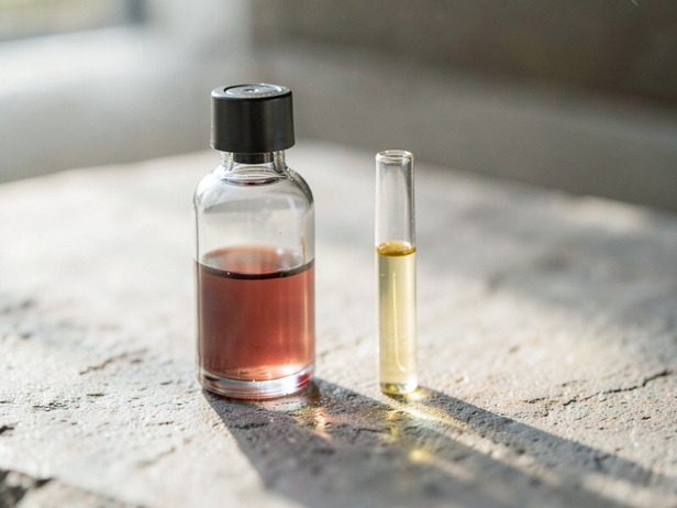
I examine a blood collection vial and centrifuge tube, showcasing a pale amber serum layer. This visual relates to understanding pet blood test reports.
The Biochemistry Panel: Organ Function in Numbers
The second section of the printout is the serum biochemistry, which measures what the organs are releasing, retaining, or failing to process.
Alanine Aminotransferase (ALT) is the most liver-specific marker in dogs and cats. It is an enzyme that lives inside liver cells; when those cells are damaged or inflamed, ALT leaks into the bloodstream. A mildly elevated ALT, one to three times the upper reference limit, can come from many things including fatty liver, certain medications, or recent vigorous exercise. An ALT running five to ten times or higher is a different conversation. Alkaline Phosphatase (ALP or ALKP) is less specific; it rises with liver disease but also with bone turnover, certain steroid use, and Cushing's disease in dogs. In cats, even a small ALP elevation carries more weight because the feline liver produces less of it baseline.
Blood Urea Nitrogen (BUN) and Creatinine together describe kidney filtration. Both are waste products the kidneys clear from blood. When they rise together, filtration is compromised. BUN can also rise from a high-protein diet, dehydration, or gastrointestinal bleeding without any kidney disease at all, which is why creatinine, less influenced by diet, matters alongside it. SDMA (Symmetric Dimethylarginine) appears on newer panels and is the earliest kidney marker currently available, detecting filtration loss before creatinine rises. Not every Indian lab offers it, but it is appearing more frequently in urban veterinary hospitals.
Glucose is measured fasting in most panels. Elevated fasting glucose in a dog or cat, confirmed on a second sample, is the primary diagnostic finding for diabetes mellitus. Cats are particularly prone to stress hyperglycaemia, a single high reading in a frightened cat at a clinic means little without a fructosamine test, which averages glucose over the preceding two to three weeks. Low glucose (hypoglycaemia) in a young puppy or kitten is a medical emergency and in an adult animal raises questions about an insulin-secreting tumour or severe liver failure.
Total Protein, Albumin, and Globulin describe the protein balance in blood. Albumin is made by the liver; a low albumin points toward liver failure, protein-losing kidney disease, or protein-losing enteropathy. Globulins include antibodies and other immune proteins; elevated globulins with low albumin in an Indian dog with a tick history is a classic pattern for chronic Ehrlichiosis. The Albumin:Globulin ratio, printed on many Indian panels, flips below 1.0 in that scenario.
Electrolytes and the Values That Often Get Skipped
Sodium, Potassium, Chloride, and Bicarbonate appear on extended panels and are sometimes skipped over because they look unremarkable. They rarely are unremarkable in a sick animal.
Potassium is the one to watch in cats with urinary obstruction, a blocked male cat can have a potassium level high enough to stop the heart before the obstruction is relieved. In a vomiting dog, sodium and chloride fall while potassium may rise or fall depending on the cause. Hypoadrenocorticism (Addison's disease) produces a classic sodium:potassium ratio below 27:1, a pattern that can be read directly off the panel numbers.
Calcium and Phosphorus matter together. Elevated calcium in a dog, particularly alongside a low phosphorus, is one of the more urgent findings on a routine panel, hypercalcaemia has a differential list that includes lymphoma, anal sac adenocarcinoma, and primary hyperparathyroidism. In a young growing animal, the ratio between calcium and phosphorus in the diet affects bone development; this is biologically relevant to the large-breed puppy owners who feed home-cooked diets without supplementation.
Amylase and Lipase measure pancreatic enzyme leakage. Spec cPL and Spec fPL, species-specific pancreatic lipase tests, are more sensitive and specific than the older amylase/lipase pair, and Indian labs in metro cities increasingly offer them. A dog brought in for vomiting and abdominal pain with a markedly elevated Spec cPL has pancreatitis until proven otherwise.
![Colorful Specimen Vials]()
Colorful Specimen Vials
Image credit : AI
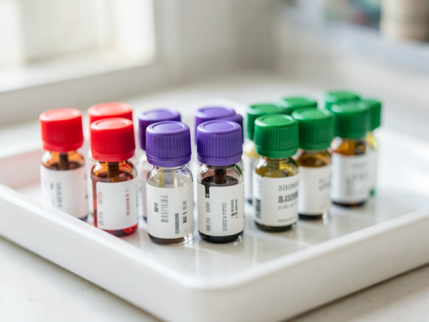
I'm examining these colorful specimen vials, a crucial step in understanding pet blood test results.
How to Read the Reference Range Column
Every Indian lab printout prints a reference range beside each value. That range is not a pass-fail line. It is a statistical description of where 95 percent of healthy animals of that species fall, which means 5 percent of healthy animals fall outside it by definition, and a sick animal can sit comfortably within it while something is going wrong.
The range also varies by species, breed, age, and sex in ways the printed column cannot always capture. Greyhounds and Salukis, kept in India as racing and coursing dogs, run higher red cell values than the standard canine range. Neonates have different glucose and protein norms than adults. A value at the very top of normal in an animal whose previous panel showed it at the bottom of normal represents a significant change, the trend matters as much as the absolute number, which is why keeping copies of every report matters.
Indian labs may use different units from those in Western reference texts. Creatinine may appear as mg/dL or µmol/L depending on the analyser. ALT might be reported as U/L or IU/L. The numbers are not interchangeable across unit systems, and a value that looks alarming in one unit may be normal in another. If the unit column is missing from your printout, ask the lab which system they use before comparing to anything you read online.
The blood report is a photograph of one moment. A single elevated ALT with a healthy, eating, active dog behind it is a reason to recheck in three to four weeks, not a diagnosis. A pattern across two or three panels over time tells a different story than any single result. The numbers gain meaning in context, the animal's age, breed, clinical signs, vaccination history, tick exposure, diet, and medication history all sit behind the values on the page, and your vet is reading all of it at once.